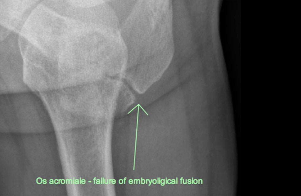
[vc_row][vc_column][mk_fancy_title color=”#000000″ size=”26″ font_weight=”200″ font_style=”normal” txt_transform=”none” margin_bottom=”30″ font_family=”none” responsive_align=”left”]In the assessment of shoulder pain, both x-rays and ultrasound should be taken.[/mk_fancy_title][vc_column_text]Without both forms of imaging it is impossible to be sure of the pathology process, and the success of treatment would be affected. MRI scan is also possible, giving some further detail deep inside the joint, but ultimately it doesn’t replace plain x-ray. An unusual finding is a so-called “os-acromiale”. This is probably not trauma, but a failure in embryology for the two parts of the shoulder blade to fuse the tip of the shoulder to the shoulder blade. The front corner of the bone then “bends” downwards and creates painful pressure on the rotator cuff, eventually causing the rotator cuff to tear.[/vc_column_text][vc_column_text]The x-ray series includes:
- AP with 20 degree caudal tilt & shoulder in external rotation
- AP with 20 caudal tilt and internal rotation.
- scapular lateral
- axillary lateral.
The best radiology departments do this series routinely.[/vc_column_text][mk_image src=”http://bos.inkserver.com.au/wp-content/uploads/2017/04/xray2.jpg” image_size=”full”][/vc_column][/vc_row]